It all starts with a sick, usually middle-aged or older dog. He is listless, not eating, maybe has vomiting and/or diarrhea, fever. These are symptoms that could mean any number of things but usually when he reaches the veterinarian’s examination table he has jaundice (yellow pigmentation visible in the whites of his eyes and possibly on his skin and gums).
Hospitalization is recommended to rehydrate him and provide supportive care. Blood tests point to a liver problem. Medications are given to minimize the liver’s workload, but soon an ultrasound is being discussed to image the liver and and costs may be rising quickly.
The array of blood tests can be confusing and it can be tempting to wait a few days and see how medical support works out before imaging. While this may be a fair choice depending on the patient, there is an important reason to image the liver quickly: the possibility that surgery is needed urgently.
The importance of an Ultrasound
While blood testing can point to the liver, the fact is that there are numerous diseases that can affect the liver. The liver can have an infection, cancer, scarring (cirrhosis), or any number of conditions. The more specific our diagnosis gets, the more specific treatment can be.
Ultrasound is a non-invasive way to evaluate the internal texture of the liver and gall bladder. By looking at the liver’s texture, it is possible to see a tumor and determine if removing it is possible or if it has invaded too far. Ultrasound can evaluate scarring and abscesses. Through ultrasound it is possible to guide a biopsy needle to an exact area to sample tissue should this be deemed necessary.
Ultrasound evaluates the gall bladder and bile ducts. One of the more important diseases to rule out promptly is biliary mucocoele because it is commonly a surgical emergency. If you wait a few days to see how the patient responds to general liver support, it may be too late to have surgery.
What Are The Gall Bladder And Biliary System?
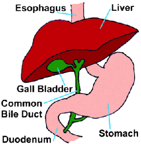 The liver serves as a toxic waste processing center for the body. It filters bacterial products (as well as nutrients) entering the body from the gastrointestinal tract and it removes toxic waste products from the bloodstream. This material that the body would like to get rid of is bound to biochemicals called bile acids. The solution of bile acids, water, mucus, pigments, and cholesterol forms the greenish yellow fluid we call bile.
Bile is made in the liver, then collected into small ducts called bile ductules and bile ducts. The bile is then moved for storage into the greenish round organ called the gall bladder. During food digestion, hormones cause the gall bladder to contract and squirt bile through the large common bile duct and into the intestine. The bile assists with digestion and carries toxins out of the body so they may be eliminated in feces. The gall bladder and its ducts represent the biliary system.
What Is a Mucocoele?
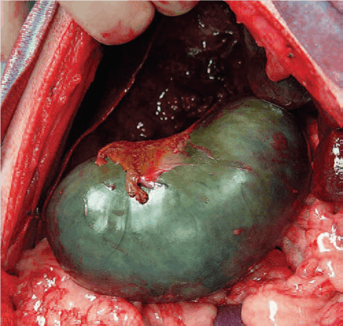
We mentioned that one of the components of bile is mucus. Normal bile is probably less than three percent mucus but when a mucocoele develops, the bile becomes mostly mucus. Normal bile is liquid but mucocele bile is thick and goopy and will not flow easily through the common bile duct. The gall bladder distends trying to pass the mucocele bile and if it actually ruptures, it is likely the patient will likely die.
The gall bladder with a mucocoele develops an appearance on ultrasound described as resembling the cut surface of a kiwi fruit. When this is seen on ultrasound in a sick, jaundiced patient, surgery to remove the diseased gall bladder should be performed as soon as possible.
The next post will be about how mucoceles form and what conditions can predispose your dog to this surgical emergency.