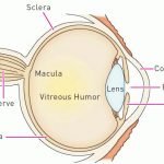
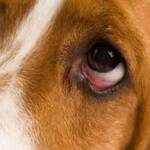
The normal lens of the eye is used to focus. It is completely clear and is suspended inside the pupil. The pupil opens and closes to control the light entering the eye so as to project an image onto the retina in the back of the eye, the way a projector projects an image onto a movie screen.
Anatomy First
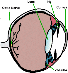
Despite being clear, tissue fibers make up the lens. As an animal ages it becomes more dense with fibers and can appear cloudy. This is called nuclear sclerosis and is responsible for the cloudy-eyed appearance of older dogs. These cloudy lenses are sometimes mistaken for cataracts, but they’re very different. Animals can see through them and the denser lens doesn’t result in any pathology.
The lens is tucked away inside it’s own capsule. A breach in the capsule would result in protein leakage into the eye and allow the body to see new material and mount an inflammatory immune response, thinking these ‘new’ proteins are foreign. The resulting inflammation (a form of uveitis) is painful and a cataract can result from this inflammation or from any of numerous other reasons listed below.
A cataract is an opacity in the lens.
Causes of Cataracts in Dogs
Cataracts can be congenital (born with it), age-related; of genetic origin (the most common cause); caused by trauma; by dietary deficiency (some kitten milk replacement formulas have been implicated); by electric shock; or by toxin. The patient with a cataract is not able to see through the opacity. If the entire lens is involved, the eye will be blind.
Many things can cause the lens to develop a cataract. One medical cause is diabetes mellitus. Diabetic cats have do not get cataracts from diabetes.
What Else Could it Be?
Many owners are not really able to tell which portion of the eye looks cloudy. Cloudiness
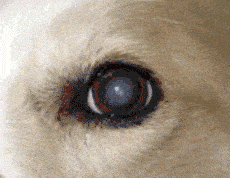
on the cornea, as caused by other eye diseases, can be mistaken for a cataract by an inexperienced owner. Also, in dogs, the lens will become cloudy with age as more and more fibers are laid down as described above. Nuclear sclerosis, as described, can mimic the appearance of a cataract, yet the eye with this condition can see and is not diseased. It is a good idea to have your veterinarian examine your pet if you think there is a cataract as you could be mistaken.
Why is it Bad to have a Cataract?
The area of the lens involved by the cataract amounts to a spot that the patient cannot see through. If the cataract involves too much of the lens, the animal may be blind in that eye and there could be cataracts in both eyes, which means the pet could be rendered completely blind.
A cataract can slip from the tissue strands that hold it in place. When this happens, the cataractous lens floats in the eye where it can cause damage. If it settles so as to block the eye’s natural fluid drainage, glaucoma (a buildup in eye pressure) can result, leading to pain and permanent blindness.
A small cataract that does not restrict vision is probably not significant. A more complete cataract may warrant treatment. Cataracts have different behavior depending on their origin. If a cataract is a type that can be expected to progress rapidly (such as the hereditary cataracts of young cocker spaniels) it may be beneficial to pursue treatment (i.e. surgical removal) when the cataract is smaller and softer, as surgery will be easier.
What Treatment is Available?
Cataract treatment generally involves surgical removal or physical dissolution of the cataract under anesthesia. This is invasive and is not typically considered unless it can restore vision or resolve pain. Pets with one normal eye and one cataractous eye can still see with their good eye and probably do not need surgery unless pain warrants it.
Owners that elect to have a cataract removed are referred to a veterinary ophthalmologist and should be prepared for the expense involved as well as the management afterward, which will include eye drops many times a day along with veterinary rechecks.
Phacoemulsification and Surgical Removal
Phacoemulsification is the most common method of removing cataracts. Here, the lens is broken apart by sound waves and sucked out with a gadget similar to a small vacuum-cleaner.
In either case, the patient is given a drug to induce complete paralysis during the surgery so as to prevent any blinking or eye movement. The patient is mechanically ventilated as the paralytic drug renders individuals unable to breathe without assistance. After the lens is removed, an artificial lens is implanted. (Without the prosthesis, the dog’s vision will be approximately 20/800, and objects will appear to be reversed, as in a mirror.)
Urban Animal Veterinary Hospital
1327 Yale St
Houston, TX 77008
(713) 863-0088
