
Most people know about melanoma and know it is a skin cancer. It arises from pigmented skin from a pigmented cell type called melanocytes. In people melanomas are commonly associated with sun exposure and overexposure.
Likewise, the situation in pets involves cancer of the melanocytes and pigmented growths. the difference is, in pets, specific body parts are classically involved.
While you should tell your family veterinarian about any pigmented growth, there are specific areas on the body to be particularly concerned about. UV rays from the sun is less important.
Benign Vs. Malignant Melanomas/Haired Skin Melanomas
The behavior of a melanoma is highly dependent on where in the body it develops. Most areas of the skin grow benign versions of melanoma that are called melanocytomas.
This means that a pigmented growth found on haired skin in an area that is not considered a danger zone is likely to behave benignly. That said, they do not all behave benignly, it is important to have all tumors analyzed by a pathologist so that a given tumor’s behavior can be predicted.
What ancillary treatment or testing is recommended (if any) will be determined based on this tissue analysis.
Many melanomas removed from haired skin show cells that look malignant under the microscope but do not behave in a malignant manner.
To get a better sense of how a given tumor will behave, the number of cells in the process of division is counted in the sample.
Often a veterinary pathologist will look at samples and use a number of criteria to determine if a mass is benign or is concerning for malignancy.
There is some suspicion that haired skin melanomas developing within 1 cm of a mucosal margin (like the mouth or genital area) behave more aggressively.
It is not wrong to have any growths sent out for pathology. In some cases, your veterinarian may refer you to a veterinary oncologist for further consult and treatment recommendations.
Below are some areas that lead toward more aggressive behavior from melanomas:
- The mouth, lips, and oral cavity
- The toe or foot
- The eye.
Each of these three scenarios is reviewed in more detail below:
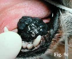
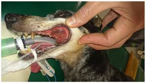
Melanoma of the Mouth
Oral melanomas are very malignant. It is locally destructive in the mouth plus it readily and quickly spreads to other areas of the body. This means that there are three aspects of the cancer requiring attention: the local destruction; tumor cells that are spreading; along with the cancer detected in other organs besides the primary site of origination.
Once a melanoma is confirmed, your veterinarian will stage the tumor to see how far it has spread (or not). Chest radiographs are used to screen metastasis to the lungs and local lymph nodes are sampled. In addition, an abdominal ultrasound is recommended to look for masses in the abdomen.
Size Matters!
In the mouth, the size of the tumor is extremely important when considering the prognosis. Veterinary medicine has adopted the World Health Organization staging system.
Stage I disease is a tumor less than 2 cm (just less than 1 inch) in diameter; Stage II disease is a tumor 2 – 4 cm in diameter; and Stage III disease consists of tumors 4 cm or larger or any tumor with local lymph node involvement. Stage IV disease includes any tumor with evidence of distant spread. Median survival times for oral melanoma have been reported as:
- Stage I: approximately 17-18 months (with surgery alone)
- Stage II: approximately 6 months (with surgery alone)
- Stage III: approximately 3 months (with surgery alone)
- Stage IV: approximately 1 month (surgery not warranted)
Additional therapies The goal of additional therapies such as radiation, chemotherapy, immunotherapy is to extend this time. In veterinary medicine, treatment is often not curative but can increase survival times.
Toe melanoma
Developing melanoma in the toe or toenail bed seems to be a particular problem for dark colored dogs. The tumor is particularly destructive to the bone and quite painful, starting usually as a swelling that seems associated with an infected toenail.
The toe infection may improve with treatment but ultimately, the issue doesn’t resolve. If the tumor has not spread beyond the toe, amputation should be curative. Unfortunately, often by the time the diagnosis is made, many of these tumors have already spread.
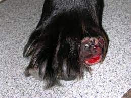
Because this is such an aggressive tumor, staging is important after melanoma is confirmed. This means chest radiographs, lymph node sampling, and ultrasound of the abdominal organs to identify distant spread.
Ocular (Eye) Melanoma
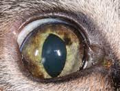
Uveal/Iris Melanoma – Dogs
The pigments of the iris and ciliary body within the eye are especially vulnerable to melanoma development. These tumors are particularly difficult to see until they are fairly large. The clinical situation is different depending on whether the patient is a cat or a dog.
The good news is that this is a benign condition in at least 80 percent of patients. In cases where progressive growth is seen, it is probably best to simply remove the eye (enucleation) though smaller growths might be amenable to laser therapy.
Uveal/Iris Melanoma – Cats
The feline situation is different. In cats, the majority of ocular melanomas are malignant and will spread. Tumors may start as flat areas of pigment on the iris (colored part of the eye) and progress to a 3D bulge. The eye will likely need removal to manage the pain from the expanding growth.
Always remember, it is best practice to stage with x-rays, local lymph node sampling, ultrasound of the abdominal organs to ensure the tumor hasn’t spread.
Iris melanosis, which is a benign condition, can be impossible to distinguish from an early melanoma so often periodic evaluation by a specialist is in order. Iris melanosis refers to the “freckles” and flat dark spots that older cats develop on their irises.
These spots are common but should not have any bulging. Often a special light on an ophthalmoscope called a slit lamp can show if the spot is starting to bulge as the slit light will warp over a 3D surface.
Epibulbar Melanoma
This form of melanoma is usually benign in either dogs or cats and arises on the outer eye portion where the sclera (white part) meets the cornea (clear part over the iris).
Smaller growths may not need treatment. Larger ones can be surgically removed, treated with laser or with cryosurgery.
Tumor spread is not expected with this form of melanoma, but it is always good practice to review histopathology and conduct chest x-rays.
Treatment
Treatment can be broken down to local control with surgery and distant control. Surgery and radiation are the fundamental treatments for local disease whereas chemotherapy is the foundation of distant disease control.
Different medications and protocols are being developed all the time. Over the last decade, a melanoma vaccine has been developed and can be helpful in management..
The Melanoma Vaccine
The melanoma vaccine has been in use for nearly a decade now, not to prevent melanoma development but to generate an active immune response against an existing tumor. The vaccine was tested on dogs with Stage II and Stage III oral melanoma after all the tumor was surgically removed.
The vaccine has resulted in longer survival times then what was found from surgery alone. The vaccine has also been found helpful for toe/digit melanomas as well.
The vaccine is for dogs without obvious visible tumor; it will probably not be helpful for dogs where there is already distant tumor spread.
Depending on the type, location and stage, your veterinarian may treat or perform surgery and chest x-rays but may recommend follow up with a veterinary oncologist to make sure your companion is getting the latest recommendations and treatment options.
If your dog or cat has any mass or growth you are concerned about, please have the area evaluated by your family veterinarian.