What is an Aural Hematoma and is it a Problem?
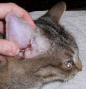
Any hematoma refers to swelling outside a body cavity, usually under the skin from a broken and bleeding blood vessel. When this occurs to the flaps of a cat or dog’s ear, it is called an aural hematoma.
Our companion animals get this often from excessive head shaking, which causes the cartilage of the ear and its vessels to break. The ear flap may partially or completely swell with blood. If the swelling extends along the length of the ear flap, it can block the opening to the ear canal.
The swollen ear flap may be uncomfortable for your animal companion and cause him/her to head shake more or paw at the ear, making the hematoma worse. While aural hematomas are more common in dogs, cats can also get them. To pet owners, the ear flap will swell and feel like a water balloon.
A very small hematoma may not initially be a problem as long as there isn’t an ear infection or another condition that can get worse and cause your pet to shake their heads.
However, if the hematoma moves toward the ear canal opening, this may create long term problems for your pet as it blocks air flow and allows dirt, debris, bacteria and yeast to build up in the obstructed canal.
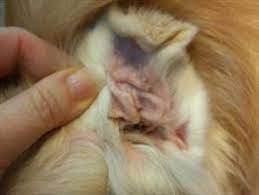
Again, a hematoma just at the ear tip will heal and scar down in a sort of wrinkled cauliflower appearance. In some cases, diagnosing and treating the underlying conditions such as allergies or an infection may stop the head shaking and a course of corticosteroids may allow the inflammation to reduce enough for the hematoma to ultimately resolve.
BUT if it doesn’t, here are the situations that require surgery to repair the damaged cartilage and stop the bleeding:
- The hematoma is so big that the ear canal is occluded (blocked). If this is the case, the ear cannot be evaluated for infection nor can any infection be treated. In this situation, the hematoma must be relieved before the ear canal can be accessed.
- The hematoma is in a location where natural healing will create scarring in such a way that the ear canal will be permanently narrowed. A permanently narrow ear canal can predispose the patient to a lifetime of ear infections. This is particularly a problem in cats.
. - The hematoma should be repaired if the owner feels the heavy ear flap is unacceptably uncomfortable for the pet.
The last reason is more for aesthetic appearances and not for medical reasons. A scarred healed hematoma may be undesirable to the pet owner. This is a little more controversial and should be a discussion with your veterinarian.

How We Treat Hematomas?
There are a handful of ways to correct ear hematomas. Some veterinarians have good results using a corticosteroid and treating any underlying ear infections or allergies, if present. But some patients fail this.
Other options include:
Aspiration
This procedure involves simply using a syringe to remove the fluid contents from the hematoma. The problem is that if the ear hasn’t healed yet, it may refill quickly when the fluid is removed.
In some cases, doing this for immediate help and trying a course of steroids and treating any ear infection may be enough, but some cases are refractory and may need surgical repair.
Sutures / Buttons – bringing the cartilage together
For this procedure, an incision is made in the ear flap surgically. The hematoma is drained of fluid and blood clots. To prevent the hematoma from refilling with fluid, multiple sutures or in some cases buttons are placed through the full thickness of the flap holding it close together.
Often bandages or some type of device is used to keep the affected ear up and close to the head, so shaking can’t occur.
Sutures are generally left in place for two to three weeks to allow scarring to take place. Because the flap is being helped together, the space for filling is closed, allowing the ear to heal and not refill.
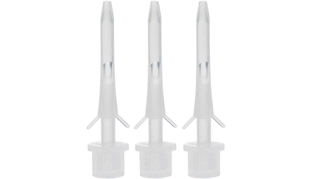
Teat Cannula Placement

A teat cannula is a small device used in the treatment of udder inflammation in cattle. But we can use it for other things, too. In the case of aural hematomas, it can be placed under sedation in a dog’s aural hematoma.
This allows the hematoma to be drained of fluids and allowed to heal over the next several weeks. This method is generally successful but does involve the dog tolerating the cannula as well as the draining of blood inside your home.
What if there is also an ear infection?
Often there is an underlying cause for a cat or dog to shake their heads. Allergies or ear infections are two common reasons. Whichever the cause, it needs to be treated at the same time as the hematoma is addressed, or else issues will keep reoccurring.
What if we leave it alone?
If left alone, an ear hematoma will eventually resolve but the area it covered may scar down and have a crumpled appearance.
Resolution of a large hematoma can take several months and be very uncomfortable for your pet.
Leaving a large hematoma alone or managing medically or in a way that is minimally invasive may be appropriate for a patient that is a high anesthetic risk or if surgery is cost prohibitive.
Hematomas in Cats

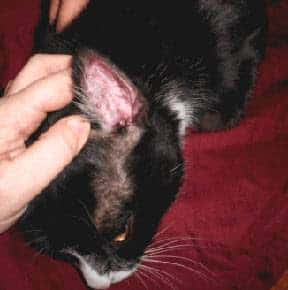
The situation in cats is somewhat more complicated than in dogs largely because the cartilage in the feline ear is more sensitive to inflammation and scarring is more severe.
This makes the untreated hematoma more likely to form a permanently narrowed ear canal, which leads to the development of life-long issues with ear infections.
If left untreated, the cat ear will often scar more severely than a dogs, so managing the hematoma and the underlying cause is very important.


