What is it?
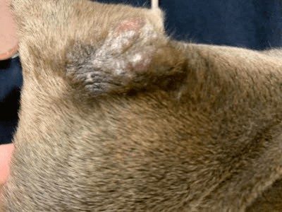
This is an interesting disease caused by a species of mycobacteria found more commonly in certain short-coated breeds of dogs such as the pitbull terrier, boxers and dobermans to name a few. This disease affects the skin of dogs causing raised bumps that are sometimes red or ulcerated and sometimes just raised with fur loss. the most common sites are the head and ears, but may be anywhere on the body. Affected dogs are otherwise healthy and are not systemically ill.
The cause and spread of the disease are not fully understood at this time but an environmental mycobacterium (such as found in soil, water) is thought to be inoculated into the skin by biting insects or poetntialy other methods.
This disease, referred to often as canine leprosy, is the most common mycobacterial disease of dogs in Australia, it has also been reported in New Zealand, Brazil, Zimbabwe, California and Florida. Disease incidence is highest in short-coated breed dogs. The disease has also been seen in other areas of the US.
What’s seen
Single to multiple skin nodule(s) that range in size from 2 mm to 5 cm in diameter. The lesions are nonpainful and don’t itch unless affected with a infection. They can become ulcerated if very large.
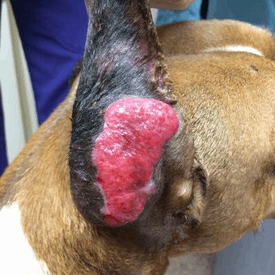
the picture above
The good news, is that the prognosis is excellent! Most cases will spontaneously regress in 3-6 weeks. If they do not resolve and /or get worse, they can be surgically removed or combination anitbiotic treatment that typically lasts for 6-8 weeks can be started.
Other things that may look like canine leprosy:
Other bacterial and deep fungal infections can cause similat looking lesions. Vasculitis, an issue affecting blood vessels to the ear may also be confused for the disease. There are also non-infectious causes for ear bumps such as suture material left behind from ear cropping. And we also can’t forget about canine cancer.
So, how do we make the diagnosis?
1. Cytology (fine-needle aspirate of nodules) with special acid-fast staining (shows mycobacterium species.
2. Dermatohistopathology – This long word means that biopsies are taken and sent for a pathology review by a pathologist specializing in skin lesions. Acid-fast staining is still used to help confirm the presence of mycobacteria.
4. Polymerase chain reaction technique on a skin biopsy (called PCR). This is amplification of DNA and can detect particular mycobacterial DNA sequences specific to dogs.
5. Mycobacterial culture – This doesn’t work as it does for other bacterial organisms because growth requirements for this bug have not yet been determined.
TREATMENT:
1. Usually a self-limiting disease, with lesions typically regressing spontaneously within 3-4 weeks.
2. If lesions persist and are few in number, aggressive surgical excision is the treatment of choice.
3. For severe, refractory, chronic, disfiguring lesions, the medical treatment of choice is a combination of antibiotics for about 6-8 weeks – until lesions have resolved or surgical removal.
After the disease has cleared, sometimes small dark colored scars are left behind. If you pooch has been diagnosed with this disease, don’t be too alarmed as it is is not considered contagious to other animals or to people.