A malignant melanoma is a tumor involving the pigmented cells of the skin. Melanoma is a classical skin cancer of humans, and in humans it is associated with sun exposure. While a good fur coat generally protects our pets from sun-induced malignant melanoma, a melanoma diagnosis is still just as serious and potentially deadly in our pets. Most patients are middle-aged or older dogs.
The melanoma is a tumor of pigmented cells called melanocytes.
Benign versus Malignant Melanomas
- The mouth, lips, oral cavity
- The toe or foot
- The eye
- Haired skin
Each of these areas produces a somewhat different syndrome and we will review each separately.
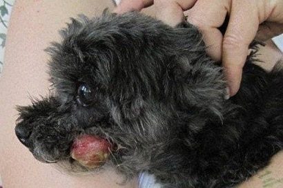
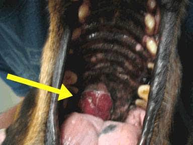
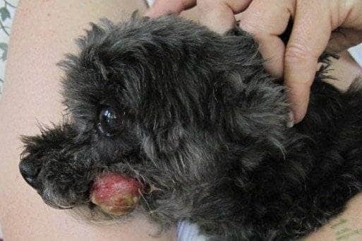
Melanoma of the Mouth
The oral melanoma is a highly malignant tumor. It is locally destructive in the mouth plus it readily and quickly spreads to other areas of the body. This means that there are three aspects of the cancer requiring attention: the local destruction in the oral cavity; undetectable tumor cells in the process of spreading; and the cancer that is detectable in distant organs.
Once a melanoma is confirmed by biopsy, a staging process must begin to determine the extent of tumor spread. Chest radiographs are scrutinized for evidence of spread, local lymph nodes are sampled, and the abdomen is screened with an ultrasound exam.
In the mouth, the size of the tumor is extremely important when considering the prognosis. Veterinary medicine has adopted the World Health Organization staging system, where Stage I disease is a tumor less than 2 cm (just less than 1 inch) in diameter; Stage II disease is a tumor 2 – 4 cm in diameter; and Stage III disease consists of tumors 4 cm or larger or any tumor with local lymph node involvement. Stage IV disease includes any tumor with evidence of distant spread. Median survival times for oral melanoma have been reported as:
- Stage I: approximately 17-18 months (with surgery alone)
- Stage II: approximately 6 months (with surgery alone)
- Stage III: approximately 3 months (with surgery alone)
- Stage IV: approximately 1 month (surgery not applicable)
The goal of additional adjunctive therapy (radiation, chemotherapy, immunotherapy) is to extend this time.
Local Disease Control
Local disease control refers to control of the disease in the mouth. Ideally most of the tumor can be surgically removed; however, even an extensive resection is not likely to remove the entire tumor with certainty and some kind of adjunctive treatment is prudent. Radiation in the area has extended the time until the tumor regrows in areas where complete margins cannot be obtained (i.e. biopsy shows there is still more tumor left). If margins are clean, immunotherapy (see later) may be all that is recommended.
Distant Disease Control
If there is documentation that the tumor has escaped and colonized another site in the body (i.e. the patient is in Stage IV), more extensive treatment is needed and this usually means chemotherapy. The melanoma is notoriously not responsive to chemotherapy in people, but research and new drug development continues. In one study carboplatin combined with piroxicam increased median survival time from 30 days to 119 days in Stage IV patients.
Microscopic Disease
After no disease is detectable to the eye, we cannot be sure about undetectable disease, which is malignant cells in transit looking for a place to set up shop. Chemotherapy helps reach these cells as does immunotherapy, a newer mode of cancer treatment. Immunotherapy involves generating an immune-response against the tumor cells and attacking them with the body’s own natural system. Periodic screening tests/staging are needed to see if the tumor has succeeded in settling in a distant organ and therapy can be ramped up to address this event.
Melanoma of the Digit (Toe)
Developing melanoma in the toe or toenail bed seems to be a particular problem for black dogs. The tumor is particularly destructive to the bone and quite painful, starting usually as a swelling that seems associated with an infected toenail. The toe infection may improve with treatment but the swelling does not resolve and ultimately gets worse. If the tumor has not spread beyond the toe, amputation should theoretically be curative but reality is that median survival time after toe amputation in cases where further tumor is not detectable is approximately one year. It is believed that 30-40 percent of these tumors have already spread at the time of diagnosis so if a longer survival is the goal, further treatment should be explored.
Because this is such an aggressive tumor, staging is important after melanoma is confirmed. This means chest radiographs, lymph node sampling, and ultrasound of the abdominal organs to identify distant spread. As before, local disease, distant disease and microscopic disease must be addressed.
Haired Skin Melanoma
The behavior of a melanoma is highly dependent on where it develops. Most areas of skin grow benign versions of the melanoma that are called melanocytomas. These tumors typically do not spread and do not behave in a malignant manner. Since some haired skin melanomas are definitely malignant in behavior, it is important to have all removed tumors (pigmented or not) analyzed by a pathologist. There is some suspicion that haired skin melanomas developing within 1 cm of a mucosal margin (like the mouth) behave more malignantly than one would expect based on what is seen under the microscope.
Many melanomas removed from the body look malignant under the microscope but do not behave in a malignant manner. To get a better sense of how a given tumor will behave, the number of cells in the process of division are counted in the sample. This is reflected by a number called the mitotic index of the tumor and is expressed as the number of mitotic figures per high power field (microscopic view). Less than three mitotic figures per high power field indicates the tumor will behave in a benign manner. There is evidence that staining the sample for certain tissue markers (such as Ki-67) can help predict behavior as well.
As with the other melanoma forms, malignant tumors should be staged and treated for local, distant and microscopic disease.
Melanoma of the Feline Eye
Uveal/Iris Melanoma – Dogs The pigments of the iris and ciliary body within the eye are especially vulnerable to melanoma development. These tumors are particularly difficult to see until they are fairly large. The clinical situation is different depending on whether the patient is a cat or a dog.
The good news is that this is benign condition in at least 80 percent of patients. The bad news is that an expanding growth inside the eye, even a benign growth, can cause pain and blindness. If glaucoma (increased eye pressure) has resulted or if there is deep inflammation in the eye, rapid tumor growth, or vision loss, it is probably worth simply removing the eye (enucleation) though smaller growths might be amenable to laser therapy.
Uveal/Iris Melanoma – Cats
The feline situation is different in that 60-70 percent of these tumors are malignant and will spread. Tumors can be large and bulgy as in the dog’s eye or they can be more subtle, starting as flat areas of pigment on the iris (colored part of the eye) and later gaining some 3-dimensional growth. The eye will likely need removal to manage the pain from the expanding growth but it is important to do proper staging first (radiographs of the chest, local lymph node sampling, ultrasound of the abdominal organs) to determine the extent of the existing tumor spread.
Iris melanosis, which is a benign condition, can be impossible to distinguish from an early melanoma so often periodic evaluation by a specialist is in order. Iris melanosis refers to the “freckles” and flat dark spots that older cats develop on their irises. These spots are common but should not have any bulging or changing roughness when the iris is viewed from the side.
Epibulbar Melanoma
This form of melanoma is usually benign in either dogs or cats and arises on the outer eye portion where the sclera (white part) meets the cornea (clear part over the iris). Smaller growths may not need treatment. Larger ones can be surgically removed, treated with laser or with cryosurgery. Tumor spread is not expected with this form of melanoma.
Treatment
As mentioned, treatment is divided into local disease control, distant disease control, and microscopic disease control. Surgery and radiotherapy are the fundamental treatments for local disease whereas chemotherapy is the foundation of distant disease control. Different medications and protocols are being developed all the time. Immunotherapy is a little harder to classify.
The Melanoma Vaccine
A melanoma vaccine has been in use for nearly a decade now, not to prevent melanoma development but to generate an active immune response against an existing tumor. The vaccine was tested on dogs with Stage II and Stage III oral melanoma after all detectable tumor was surgically removed with improved survival times achieved over what was expected from surgery alone. The vaccine has also been found helpful for toe/digit melanomas as well.
The vaccine is for dogs without grossly detectable tumor; it will probably not be helpful for dogs where there is already distant tumor spread.
How it Works
An enzyme called tyrosinase is crucial to the melanocyte’s ability to produce melanin (pigment). By using human tyrosinase as a stimulator, the patient’s immune system can be tricked into attacking the melanoma cells that contain the patient’s natural tyrosinase. The vaccine is available only through veterinary oncology specialists. It is given in four single doses at 2-week intervals. Booster shots are given every 6 months and is best used for dogs with oral melanoma without node involvement. Life expectancy has been extended to over one year in many cases.